Additional information
| Weight | 407 g |
|---|---|
| Dimensions | 24 × 16 cm |
| Authors | |
| ISBN | 9789062992355 |
| Publisher | |
| Publication Year | 2012 (9-11-2012) |
€25,00 excl. VAT
Also available as ebook on eBooks.com.
Get it on Google Play.
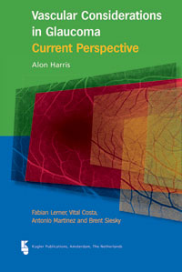
Open angle glaucoma (OAG) is one of the leading causes of impaired vision worldwide. The pathogenesis of glaucomatous optic neuropathy remains poorly understood, and several pathogenic mechanisms are proposed to coexist. As the world population ages, OAG will become more prevalent and advances in the diagnosis and treatment of glaucomatous optic neuropathy are important to protect and improve the quality of life of our aging population.
Treatment of OAG has been directed at lowering intraocular pressure (IOP) which is the only current therapeutic strategy available to patients with glaucoma. While a wide variety of studies have demonstrated that lowering IOP decreases the risk of glaucoma development and/or progression, many studies have also shown that some patients continue to lose vision despite significant lowering of IOP.
There have been many attempts to elucidate the etiology for the deterioration in glaucomatous optic neuropathy despite low levels of IOP. Over the past several decades, deficits in the ocular circulation of patients with OAG have become well established and these may explain the continued progression of OAG patients despite lowered IOP.
The purpose of the present publication is to provide an updated view of ocular blood flow and vascular dysregulation in OAG. The importance of the topic was demonstrated by the focus of the 2009 6th Consensus meeting of the World Glaucoma Association which focused entirely on blood flow deficits in patients with OAG. Although a great deal of knowledge on vascular risk factors in glaucoma has already been established, many questions remain.
Do ocular blood flow deficits precede glaucoma progression? How does ocular perfusion pressure fit into the IOP and blood flow paradigm? What conclusions can be drawn from recent evidence showing the fluctuation of OAG risk factors including IOP, blood pressure and ocular perfusion pressure?
We hope this update d current prospective will serve as a foundation for future investigations which will be designed to answer these and other important considerations in the management of glaucoma.
Alon Harris, MS, PhD, FARVO
Director of Clinical Research
Lois Letzter Professor of Ophthalmology
Professor of Cellular and Integrative Physiology
1 Background
2 Clinical measurement of ocular blood flow
2.1 Color Doppler imaging
2.2 Laser Doppler flowmetry and scanning laser flowmetry
2.3 Retinal vessel analyzer
2.4 Blue field entoptic stimulation
2.5 Laser interferometric measurement of fundus pulsation
2.6 Dynamic contour tonometry and ocular pulse amplitude
2.7 Pulsatile ocular blood flow analyzer
2.8 Laser speckle method (laser speckle flowgraphy)
2.9 Digital scanning laser ophthalmoscope angiography
2.10 Doppler optical coherence tomography
2.11 Retinal oximetry
3 Autoregulation
3.1 Mechanisms of autoregulation
3.2 Impaired autoregulation in glaucoma
3.3 Mechanisms of impairment and damage
4 Vascular risk factors
4.1 Blood pressure and perfusion pressure
4.2 Age
4.3 Migraine
4.4 Disc hemorrhage
4.5 Diabetes
4.6 Emerging risk factors
5 Diurnal variations of intraocular pressure, blood pressure and perfusion pressure
5.1 Diurnal variations of intraocular pressure
5.2 Blood pressure, perfusion pressure and glaucoma
5.3 Diurnal ocular perfusion pressure and glaucoma
6 The relationship between blood flow and structure
6.1 Capillary dropout
6.2 Blood pressure and optic disc morphology
6.3 Blood flow and optic disc morphology
6.4 Blood flow and retinal nerve fiber layer thickness
6.5 Circadian flow fluctuations and retinal nerve fiber layer
6.6 Central corneal thickness
7 Ocular blood flow and visual function
8 Cerebrospinal fluid pressure and glaucoma
9 Topical medications and ocular blood flow
9.1 Background
9.2 Topical CAIs
9.3 Topical carbonic anhydrase inhibitors and visual function
| Weight | 407 g |
|---|---|
| Dimensions | 24 × 16 cm |
| Authors | |
| ISBN | 9789062992355 |
| Publisher | |
| Publication Year | 2012 (9-11-2012) |
