Additional information
| Publication Year | 2025 |
|---|---|
| ISBN | 90-6299-297-8, 978-90-6299-297-3 |
| Pages | 170 |
| Authors | |
| Publisher |
€39,00 excl. VAT
Introduction
Preface
In Appreciation
Abbreviations
TUTORAL ONE
The Geography of the Retina
Retinal Facts and Figures
The Peripheral Fundus
The Ora Serrata
The Nasal Ora Serrata
The Temporal Ora Serrata
The Pars Plana
The Vitreous Base
Dentate Processes
Average Ora Serrata Contains: (from 200 autopsies)
THE VITREOUS
Aging Changes
Tractional Effects
Peripheral Retinal Degenerations
Classification Of Retinal Breaks
Tractional Tears
Paving Stone (Cobblestone) Degeneration
TUTORIAL TWO
The Patient and the Pupil
Use as follows
The Examination Room
TUTORIAL THREE
Essential Optics
The Elbow Optics Method
Clinical Applications
TUTORIAL FOUR
Scleral Depression
The Thimble Depressor
Modification
The Articulating Depressor
The Long Depressor
Scleral Depression
Clinical Applications
TUTORIAL FIVE
The Fundus Drawing
The Approach
Localization
Conformation
Colors
TUTORIAL SIX
The Scleral Buckle
Basic Principles
Essential Principles and Techniques
The Scleral Flap Dissection
Dissection of the Flaps
Anchor Sutures
Creating the Scleral Dissection
The Incision
The Perforation and The Perforation Fluid ‒ General Management
A Cautionary Note
ADDENDUM
Cryopexy Techniques
CONCLUSION
A lecture is a passive experience, a lecturer speaks, the student listens. Pictures are projected in a darkened room, it is early in the morning, students doze off.
So, I left the lights on. I see the eye remained open, but the minds are asleep.
Better but not Best.
The Best is to switch from lecture to seminar around a table.
The ‘Lecturer’ becomes a ‘Tutor’ who leads. All participants contribute.
This Handbook is our guide, our map for our journey.
It lies flat on the table. Participants turn each page, as we proceed. Look, read and discuss each subject, make notes.
Lights are on, no slides that dazzle, eyes open, minds awake, alert.
Understanding, to the completion of our journey together.
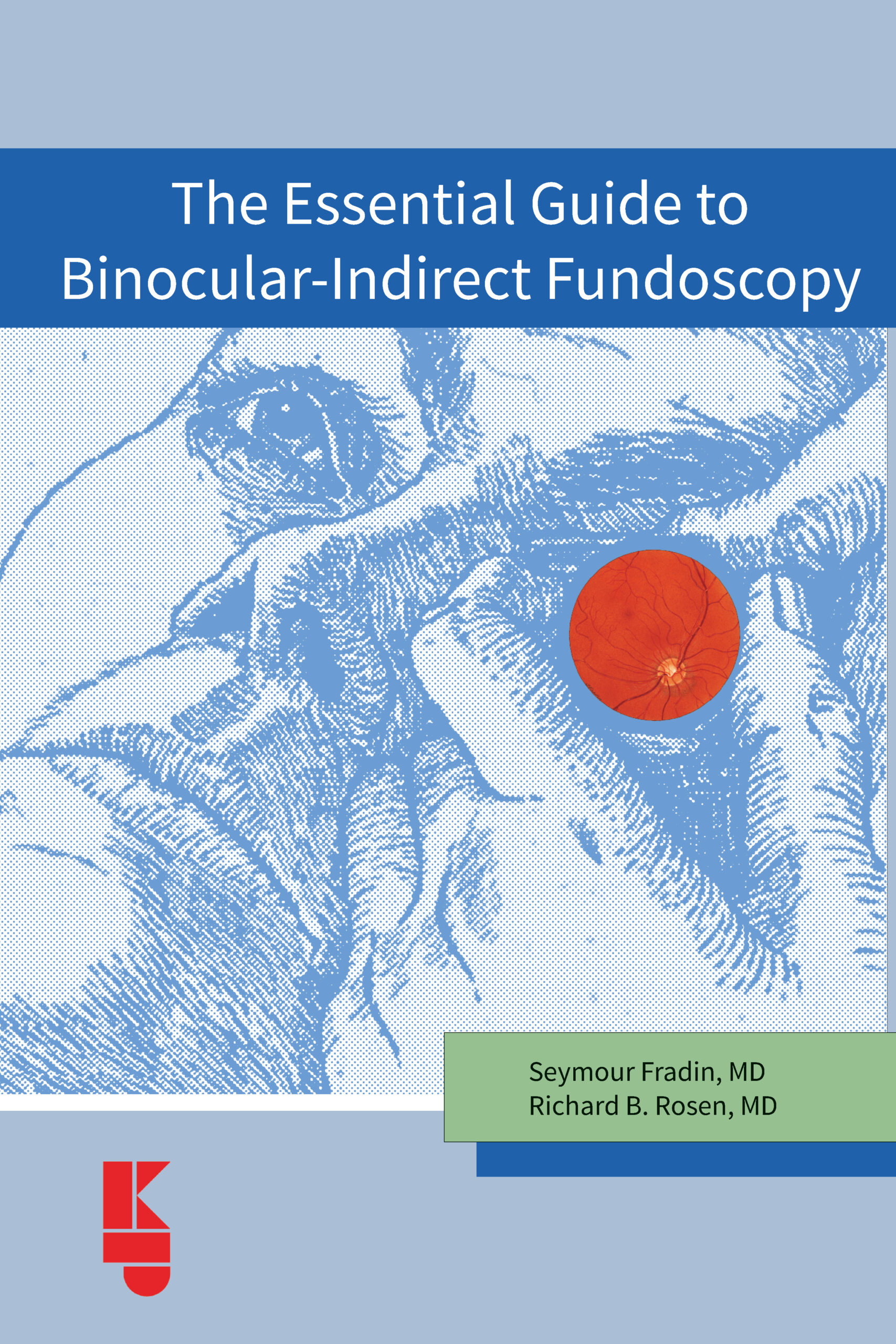
Seymour Fradin, MD
New York
Fundus examination with the indirect ophthalmoscope is among the most challenging diagnostic techniques one must master in the examination of the eye. It demands disciplined positioning, a sensitivity for the delicate subject being examined, and a fluidity of execution. Inspiring compliance in our examinee is critical for optimal peripheral retina assessment and to
avoid missing important details.
Imparting these skills to trainees has been Seymour Fradin’s passion for more than sixty years. His own experience as an World War II veteran, engineer, aviator, scientist, medical artist, retinal surgeon, and teacher have provided him with a rare combination of perspectives into the optics, mechanics, medical science, and bedside manner that few enjoy.
This handbook encapsulates the core of Seymour’s one-on-one tutorials with trainees at all levels. His illustrations reveal details that we can all aspire to gaze upon and his many pearls will help you move many steps forward toward your own mastery of the technique.
Richard B. Rosen, MD
| Publication Year | 2025 |
|---|---|
| ISBN | 90-6299-297-8, 978-90-6299-297-3 |
| Pages | 170 |
| Authors | |
| Publisher |