Additional information
| Weight | 2000 g |
|---|---|
| Dimensions | 28 × 22 cm |
| Editors | |
| ISBN | 978-90-6299-295-9 |
| Biblio | Book. 2023. Hardbound. US letter format, with many full color figures. |
| Publication Year | 2024 |
€100,00 excl. VAT
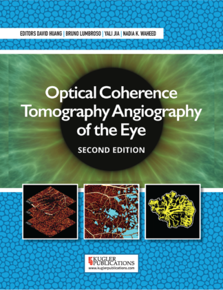
Optical coherence tomography angiography (OCTA) has undergone tremendous growth since its first commercial introduction in 2014. Because it provides injection-free, capillary-resolution, 3-dimensional angiography of the retina and choroid, OCTA is likely to overtake fluorescein as the most important angiographic imaging technique in the eye. Nearly all manufacturers of ophthalmic OCT now offer OCTA products. A PubMed search now yields over 5700 articles on OCTA and related terms.
Clinical investigators have already found a use for OCTA in almost every category of retinal and optic nerve diseases. This book is meant to bring together all this information so clinicians can have one authoritative text to turn to as we begin to use this new imaging modality that was never taught when we were in formal training.
Also available as ebook on eBooks.com.
Get it on Google Play.
Optical coherence tomography angiography (OCTA) has undergone tremendous growth since its first commercial introduction in 2014. Because it provides injection-free, capillary-resolution, 3-dimensional angiography of the retina and choroid, OCTA is likely to overtake fluorescein as the most important angiographic imaging technique in the eye. Nearly all manufacturers of ophthalmic OCT now offer OCTA products. A PubMed search now yields over 5700 articles on OCTA and related terms. Clinical investigators have already found a use for OCTA in almost every category of retinal and optic nerve diseases. This book is meant to bring together all this information so clinicians can have one authoritative text to turn to as we begin to use this new imaging modality that was never taught when we were in formal training.
Many of us have spent many hours on fluorescein rounds during our residency and fellowship to learn the principles of fluorescein angiography (FA) and the myriad manifestations of retinal diseases. Unfortunately, OCTA is sufficiently different that we need to start this educational process all over again. Let’s begin with the bases of OCTA, which are high-speed OCT systems and efficient computational algorithms. These are covered in the first 2 chapters, which the clinicians should read but not worry about understanding or retaining all the technical details. Get the rough gist so you have some basis for choosing among the available commercial systems covered in Chapters 9-15. It is useful to read up on the particular OCTA system that you are using, so you can take advantage of all the available features.
All OCTA algorithms measure changes in the OCT signal over time, from which the flow signal is derived, as well as the attendant bulk motion and flow projection artifacts. The intrinsic flow contrast comes from the motion of blood cells and does not require the injection of any dye or contrast agent. OCTA is 3-dimensional and can resolve up to 4 separate vascular layers (plexuses) in the retina. Unlike FA, which detects abnormal blood vessels by dye leakage, OCTA detects neovascularization and other abnormal vessels by their occurrence in the wrong layer, as well as their characteristic patterns. To the user, going from the 2-dimensional FA to 3-dimensional OCTA is like going from driving a car to flying an airplane – a new set of skills is needed to deal with the extra dimension. The skills needed to understand en face and cross-sectional visualizations of OCTA and recognize true pathologies versus artifacts are taught in Chapters 3-5. Chapters 3 and 5 are must-reads for most clinicians, while the visualization of anterior eye circulation discussed in Chapter 4 is more geared toward researchers in this area. Quantifying flow (Chapter 6) is important for providing endpoints for clinical studies, for example, to measure the response of choroidal neovascularization to therapy, or to monitor the progress of diabetic retinopathy or glaucoma. Some OCTA systems offer basic quantification tools, which will likely become common and more sophisticated in the next few years. Our authors also shared their pioneering work on leveraging artificial intelligence for analyzing OCTA in quantification and diagnosis (Chapter 7). Before moving on to the clinical chapters, the readers need to have a quick look on Chapter 8 , which teaches standard OCTA terminology, some of which use similar words with new specific meanings. The reader is advised to refer to this chapter as a glossary later if there is any doubt as to misunderstanding any terms in the subsequent clinical chapters.
The clinical chapters (16-46 ) were written by a wide collection of experts. We apologize that their style may vary and some content may overlap. It is, of course, impossible to read through all these chapters in one sitting. The readers are advised to use these chapters as references when they encounter questions on the interpretation of OCTA in the clinic. The most important application of OCTA at the current time is probably in the confirmation of choroidal neovascularization and the differential diagnosis of its underlying etiology. Thus, the related chapters are of particular importance. Macular telangiectasia (Chapter 22) and central serous chorioretinopathy (Chapter 23) are less frequently encountered conditions, but are important to read about because OCTA offers new information that can improve the staging and management of these diseases. Finally, glaucoma specialists should become familiar with the reduction in capillary density in the optic nerve head, peripapillary retina, and macula that occurs in glaucoma of all varieties. As quantification tools improve, this is likely to become an important tool for diagnosing and monitoring this leading cause of blindness worldwide.
Because OCTA is noninvasive, rapid, low cost, and easily available as a software add-on to the current generation of high-speed OCT systems, it can be used at every visit to screen for suspected diseases and every follow-up visit to monitor treatment efficacy. It is likely to be used far more than FA ever was. It is worth investing the time to learn about this new imaging modality now, and to closely follow the rapid expansion of knowledge on how to interpret and use OCTA in everyday clinical practice.
| Weight | 2000 g |
|---|---|
| Dimensions | 28 × 22 cm |
| Editors | |
| ISBN | 978-90-6299-295-9 |
| Biblio | Book. 2023. Hardbound. US letter format, with many full color figures. |
| Publication Year | 2024 |
